Description
Bone Health:
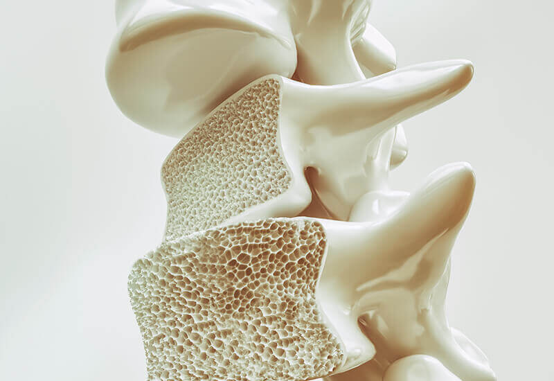
Strong, healthy bones are important for providing structure and anchoring muscles and ligaments, protecting our internal organs and storing essential minerals. During childhood, adolescence and as young adults, new bone is produced more quickly than old bone is broken down, until reaching a peak bone mass at around 30 years of age. Bone remodeling continues throughout adulthood, but bone mass slowly decreases, as the old bone removal (resorption) is quicker than new bone production (ossification). In many elderly people, this bone mass loss results in osteoporosis, a condition characterized by thin, porous and brittle bones, and an increased risk of bone fracture. Osteoarthritis is another health issue that frequently affects elderly people. Osteoarthritis is the most common form of arthritis and occurs when the protective cartilage within the joints wears down.
TURNAROUND TIME
SAMPLE TYPE
AGE REQUIREMENT
GENDER
Test Details
What factors affect bone health?
There are several factors that contribute to bone health, including gender, hormone levels, ethnicity, physical activity, medications, nutrient uptake and genetics.
Women have an increased risk of osteoporosis, due to a lower bone mass and decreased estrogen levels that occur at menopause. Variations in male hormones also influence the risk, as men with lower testosterone levels can have a lower bone mass. Hispanics and African-Americans tend to have higher bone densities and a reduced risk of osteoporosis compared to Caucasians and Asians. Weight-bearing exercises (e.g. running, walking and dancing) help to strengthen bones, hence a sedentary lifestyle increases the risk of poor bone health. The long-term use of many medications (e.g. corticosteroids) can also reduce bone mass.
Strong bones require adequate calcium in the diet. Vitamin D is also essential, as the calcium cannot be absorbed without healthy vitamin D levels. A poor diet or a decreased ability to absorb these essential nutrients increases the risk of osteoporosis. Gastrointestinal issues (e.g. Crohn’s and celiac disease) impact an individual’s ability to absorb nutrients from the diet. Lactose intolerance is also associated with a lower bone mass, as these individuals are unable to digest dairy products that are a rich source of calcium.
Genetic variation is a major factor influencing bone health. Genetic changes affect the risk for gastrointestinal issues that affect mineral absorption (e.g. HLA-DQA1 and HLA-DQB1 changes increase the risk of celiac disease and common variations in the MCM6 gene cause lactose intolerance). Variations in the CYP2R1, GC, WNT16, GDF5 and COL1A1 genes are all associated with bone and joint health issues by interrupting essential vitamin D activity and cell-to-cell signaling, inhibiting bone, joint and cartilage maintenance, and affecting collagen production. This genetic analysis identifies common variations in the CYP2R1, GC, WNT16, GDF5 and COL1A1 genes that are associated with an increased risk of osteoporosis and osteoarthritis. Genetrace also offers genetic testing for celiac disease risk and lactose intolerance.
Vitamin D
Vitamin D is an essential fat-soluble vitamin required for the absorption of other nutrients, particularly calcium and phosphate. Ultraviolet B radiation (from sunlight) triggers the synthesis of vitamin D in the body. Vitamin D can also be obtained from some foods, including fatty fish, fish liver oils and fortified foods (e.g. infant formula, milk and cereals). Decreased vitamin D levels and activity result in reduced calcium absorption and increased risk of osteoporosis.
- CYP2R1 – Vitamin D from the diet or triggered by sun exposure must be converted to the physiologically active form by a two-step process. Cytochrome P450 2R1 (encoded by the CYP2R1 gene) is the enzyme responsible for the first conversion step from vitamin D to calcidiol. Variants of this enzyme are associated with reduced enzyme activity and reduced levels of active vitamin D.
- GC – The vitamin D binding protein (encoded by the GC gene) is required to transport active vitamin D around the body and into the cells. Variations of this protein reduce the efficiency of vitamin D transport and cellular uptake.
The Wnt signaling pathway
The Wnt signaling pathway is one of the most important pathways in several biological processes, including embryonic development, postnatal development, adult tissue homeostasis and bone biology. The Wnt cascade is especially important for signaling the differentiation of mesenchymal stem cells into osteoblasts – the cells responsible for bone formation.
- WNT16 – The WNT16 gene encodes a protein from the Wnt signaling pathway. Inactivating variations in this gene disrupt the Wnt cascade, resulting in reduced osteoblasts and bone formation. This leads to decreased bone density and an increased risk of osteoporosis.
Bone, joint and cartilage maintenance
The maintenance, development and repair of bones, joints and cartilage is a lifelong process, but decreases as we age. Many elderly people suffer from osteoarthritis, which occurs when the protective cartilage breaks down, leading to joint pain, stiffness and inflammation.
- GDF5 – The GDF5 gene encodes an important regulator protein for the maintenance, development and repair of bones, joints and cartilage. A variant of this gene reduces the expression of the regulator protein and increases the risk of osteoarthritis and fractures. This variant is also associated with an increased risk of congenital hip dysplasia, which is an instability or dislocation of the hip joint in infants and young children due to a poorly formed hip joint.
Collagen
Collagen is an essential strength and structural component and the most abundant protein in mammals. It exists as a rigid form in bones, a gradient form in cartilage and a compliant form for many other tissues (e.g. skin, muscle, tendons, ligaments and blood vessels).
- COL1A1 – The COL1A1 gene encodes the major component of type I collagen, found in bone, skin and tendons. A variant of COL1A1 affects the formation of this collagen and is associated with decreased bone mineral density and osteoporosis in postmenopausal women.


